
Core instrument facilities are a key part of the University’s research infrastructure. These facilities provide shared resources that are otherwise inaccessible to many potential users in the community due to cost or expertise constraints. Many researchers may require a service only occasionally and cannot justify the cost of operating their own capability for their use alone. Moreover, our core facilities are open to users from outside the University, including local and regional institutions of higher education and industry customers who otherwise may not have local access to these advanced services.
Quick navigation:
- Access and Scheduling
- Blatt BioImaging Center
- Materials Research Core
- Syracuse University Microanalytical Laboratory
- Laboratory Animal Resources
The instruments, machines, and laboratories in core facilities are maintained and operated by expert staff with deep knowledge of the equipment and services being provided. Our experts keep the physical infrastructure of the cores in top operating condition by performing repairs or maintenance personally and by interfacing with vendor technicians as required.
The cores are user facilities, allowing student, staff, and faculty researchers hands-on access to the instruments at affordable rates. Our expert staff train users on the instruments and certify their ability to operate them independently, and staff can also assist users on experimental design and data analysis. Optionally, researchers can opt for fee-for-service, where technical staff analyze samples for a fee.
Access and Scheduling
The core facilities at Syracuse University use Facilities Billing System (FBS) to manage training, access, scheduling, invoicing, and billing. To view the cores in FBS, examine specific resources, and request access, please visit the University’s FBS portal. Each of the cores has separate access and usage procedures, so please consult individual FBS pages and their websites, linked below.
These core facilities are not an exhaustive list of equipment, shared facilities, and technical expertise at Syracuse University. For resources not in core facilities, please also see department and unit-level information.
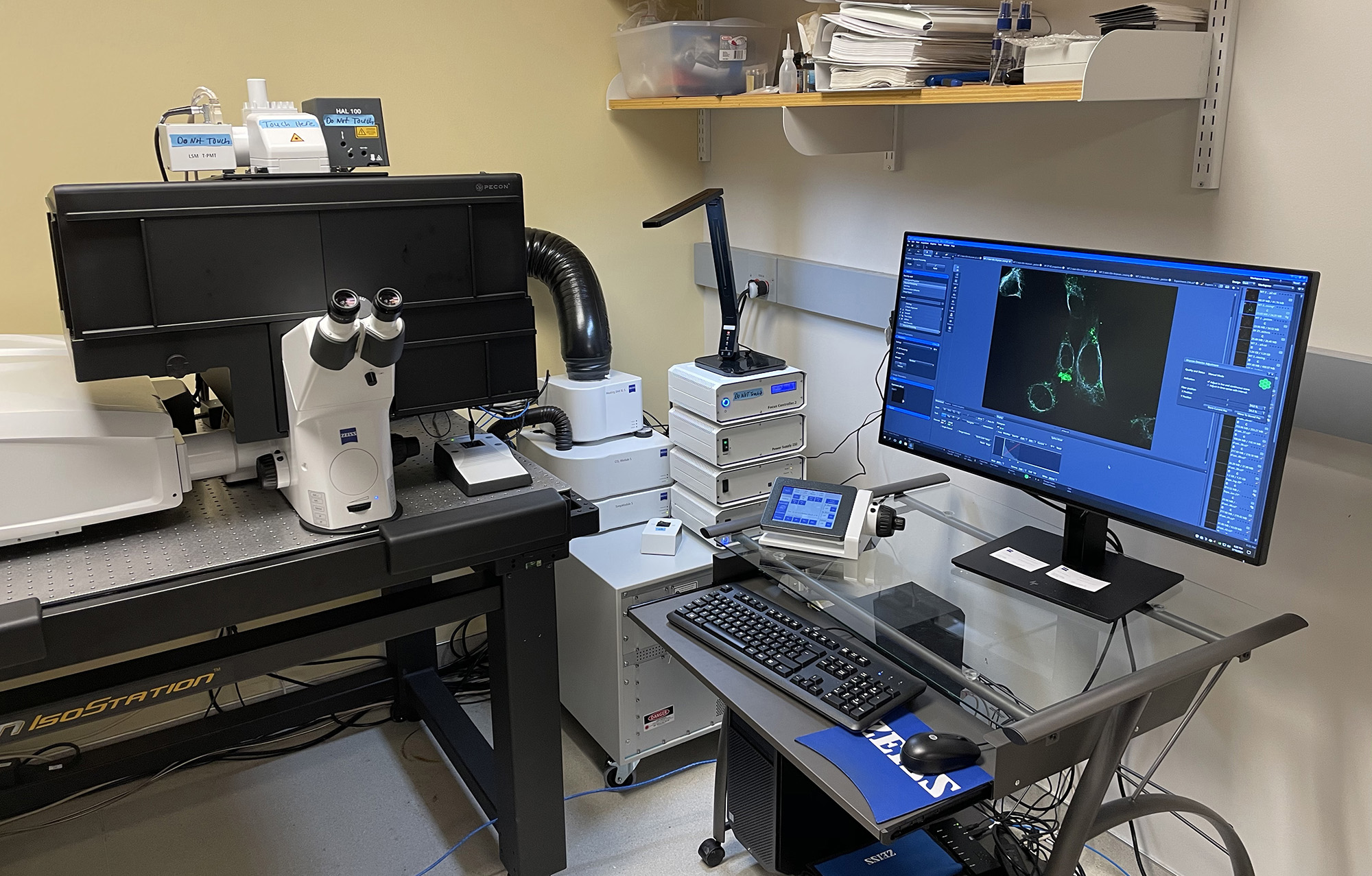
Blatt BioImaging Center
The Blatt BioImaging Center is a light microscopy core facility that focuses on live cell and enhanced resolution imaging of diverse model organisms ranging from cells to organoids and whole organisms. Located on the 2nd and 3rd floors in Life Sciences Complex (Rooms 249, 259, 359 and 318) the imaging center houses state-of-the art confocal microscopes and high-resolution bright field imaging systems. Highlights include:
- Zeiss LSM 980 with Airyscan II, super-resolution laser scanning confocal
- Leica DMi8 with digital lightsheet, laser scanning confocal
- Andor Dragonfly-620 SR, spinning disk confocal with TIRF
- Zeiss AxioObserver Z1, spinning disk confocal
- Leica Thunder systems, including 1) stereoscope with micromanipulator and 2) compound microscope for tissue/organoid
Materials Research Core
Materials Research Core Facility is located in Bowne Hall (4th Floor) and Link Hall (basement). It is a cornerstone of the research capabilities of biomedical engineering, chemistry, and the physical sciences. The facility is an enabling resource for researchers engaged in a wide spectrum of problems, ranging from fundamental studies of the biochemical and physical processes controlling cell functions to the development of new technologies for biomedical applications. Highlights include:
- Zeiss Sigma 360 field-emission scanning electron microscope with variable pressure module.
- Optical microscopy (Leica DMI4000B inverted, Hirox digital microscope)
- Thermal Analysis (TA Discovery DSC 250, TA Discovery TGA 550)
- Mechanical properties and morphological characterization (Rame-Hart 250-F1 contact goniometer, Test Resources tensile-compression tester, TA DHR3 rheometer, TA Q800 dynamic mechanical analyzer, MTS model 204 load frames)
- Molecular/Solution Characterization (Thermo iS5 FTIR with ATR, Malvern Zetasizer Ultra, BD Accuri flow cytometer, Biotek Synergy 2 plate reader)
- Materials Fabrication (Fluidnatek LE-50 electrospinner, carver digital press)
- Histology suite with Epredia tissue processor, embedder, and microtome

Syracuse University Microanalytical Laboratory
The Syracuse University Microanalytical Laboratory is a user facility located in Heroy Geology Laboratory, home of Earth & Environmental Science. The facility houses such instrumentation as an electron microprobe and Raman microscope that can be used to study solid materials such as glasses, minerals, bone, ceramics, metals, etc.—essentially any material that will not evaporate can be imaged and analyzed. Highlights include:
- Cameca SXFive electron microprobe
- Renishaw inVia confocal Raman microscope
- JEOL JCM-6000Plus benchtop SEM
- Bruker Vertex 70v Fourier transform infrared spectrometer
Laboratory Animal Resources
LAR is responsible for providing high quality animal care, veterinary care, and support for the research and teaching at Syracuse University. The highly dedicated and experienced LAR staff is committed to the health and well-being of laboratory animals as well as student and investigator training. LAR currently accommodates mice and zebrafish.